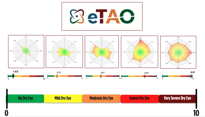
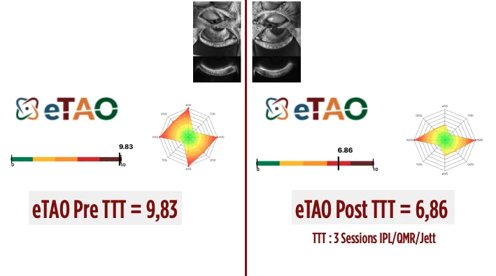
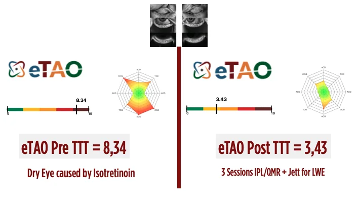
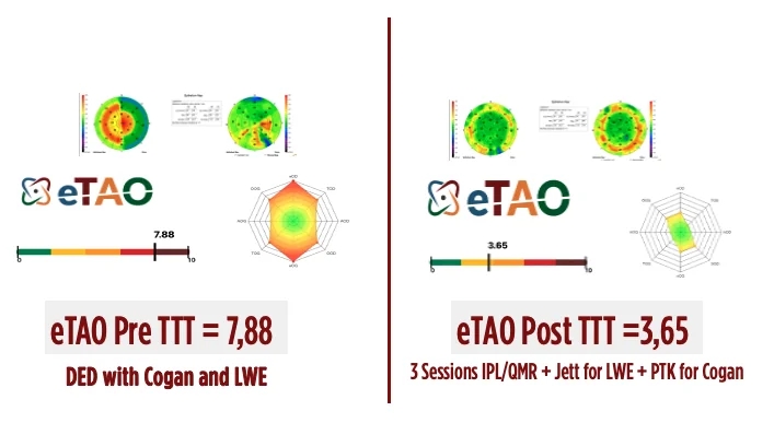
The score measures severity. The radar reveals the profile.
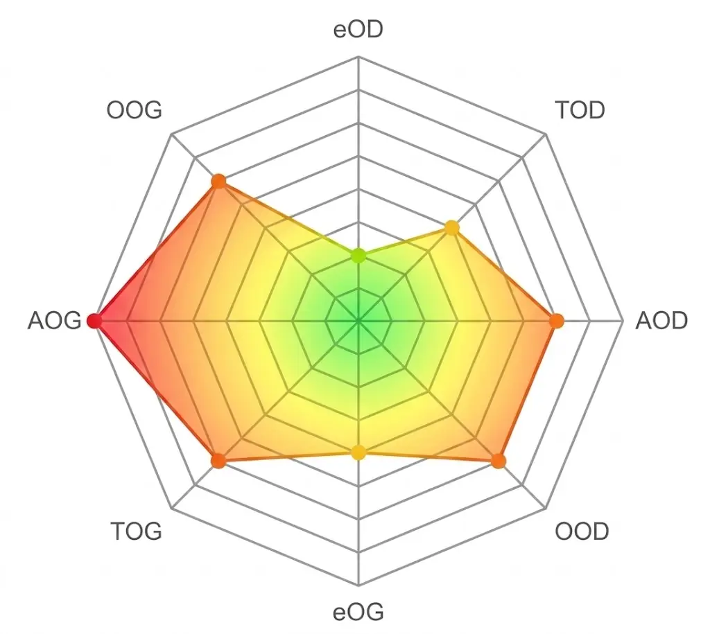
Four Clinical Dimensions
e Corneal Epithelium
The e component assesses corneal epithelial involvement.
Clinical examination with fluorescein identifies the presence, distribution and severity of epithelial staining, ranging from a normal ocular surface to localized or diffuse epithelial damage. The eTAO classification distinguishes four clinical patterns.
The Oxford staining scale provides the clinical basis for assessing corneal epithelial damage, while eTAO reorganizes these findings into four clinically relevant categories.
- e1No stainingOxford grade 0
- e2No staining but excessive tearingReflex tearing without staining
- e3Localized stainingOxford grades I, II and III
- e4Diffuse stainingOxford grades IV and V, or Cogan-like epitheliopathy
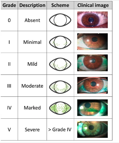
From clinical appearance to structured grading
The eTAO epithelial score distinguishes not only the severity of corneal staining, but also different clinical patterns of epithelial involvement.
This is particularly important because significant ocular surface dysfunction may be present even in the absence of conventional corneal staining: reflex tearing without staining (e2) is itself a sign of surface instability.
- e1
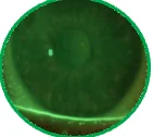 No staining
 - e2
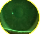
 No staining but tearingReflex tearing without epithelial damage
 - e3
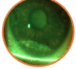 Localized staining
 - e4
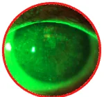
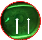 Diffuse staining and/or Cogan-like epitheliopathy
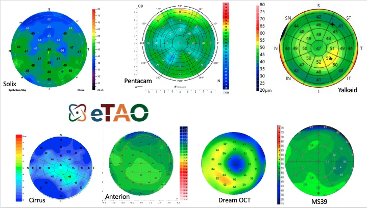
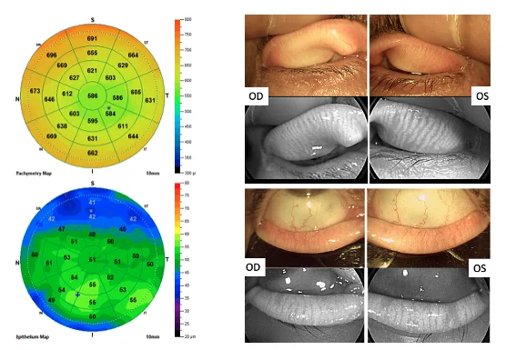
In eTAO Expert, clinical assessment can be further refined by corneal epithelial OCT mapping.
T Telangiectasia
The T component evaluates vascular involvement of the eyelids and palpebral conjunctiva.
Telangiectatic changes can progressively involve different anatomical levels.
- T1
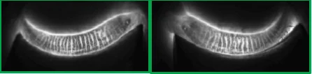 No telangiectasia
 - T2
 Lid margin or upper-eyelid telangiectasia
 - T3
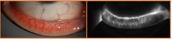 Superficial telangiectasia of the palpebral conjunctiva
 - T4
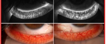 Deep telangiectasia of the palpebral conjunctiva
Assessing these vascular changes provides complementary information on the inflammatory and vascular component associated with meibomian gland dysfunction and ocular surface disease.
A Meibomian Gland Atrophy
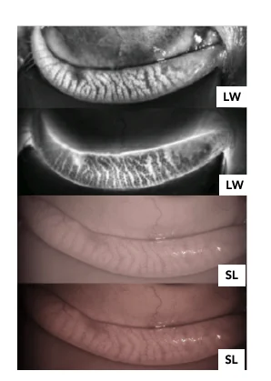
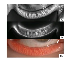
The A component provides a structural assessment of the meibomian glands.
Meibography allows gland loss to be visualized and quantified according to four levels.
- A1
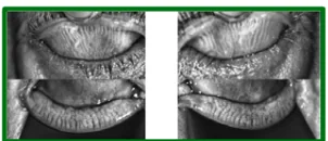 0 to 25% gland atrophy
 - A2
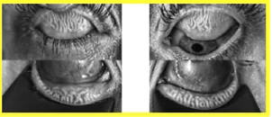 26 to 50% gland atrophy
 - A3
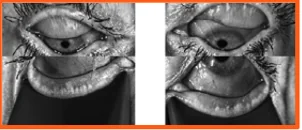 51 to 75% gland atrophy
 - A4
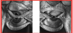 More than 75% gland atrophy
Structural gland loss provides important information on the severity and chronicity of meibomian gland dysfunction. This four-level grading is consistent with established meibography scales (meiboscore).
Whenever possible, both upper and lower eyelids should be evaluated to obtain a comprehensive assessment of meibomian gland structure.
O Meibomian Gland Obstruction
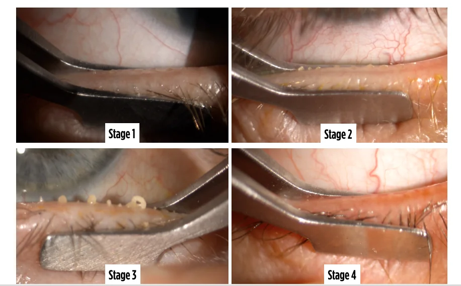
The O component evaluates meibomian gland function through gland expression and assessment of meibum quality.
Four stages are distinguished.
- O1
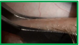 Clear, transparent meibum with easy expression
 - O2
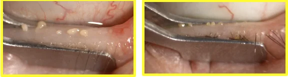 Cloudy or abnormally viscous meibum
 - O3
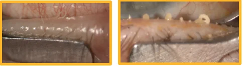 Thick, toothpaste-like or filamentous meibum
 - O4
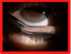 Complete obstruction with absence of expressible meibum
Unlike meibography, which assesses gland structure, the O score provides functional information about gland obstruction and secretion.
A Structure O Function
Progressive meibomian gland obstruction
The clinical appearance of expressed meibum changes progressively from clear and easily expressed secretion to thickened meibum and, ultimately, complete gland obstruction.