WIDES promotes a mechanism-based diagnostic approach.
Accurate Diagnosis for an Optimal Interventional Therapy
eTAO translates diagnostic findings into a structured, standardized profile designed to support mechanism-based and personalized dry eye management.
Symptom Assessment
OSDI-6
A standardized symptom questionnaire used as the initial step in the diagnostic evaluation of dry eye disease.
Slit-Lamp Examination
The essential first clinical assessment
A careful slit-lamp examination remains one of the most informative steps in dry eye evaluation. Before performing advanced imaging or instrumental testing, a systematic examination of the ocular surface and eyelids can already provide essential information about the predominant mechanisms involved.
With appropriate training, slit-lamp examination allows the clinician to identify and semi-quantify several key components of dry eye disease and meibomian gland dysfunction, including ocular surface staining, visible meibomian gland atrophy, telangiectatic involvement, and meibomian gland orifice obstruction.
It therefore provides an immediate clinical framework that helps determine which additional diagnostic investigations are most relevant.
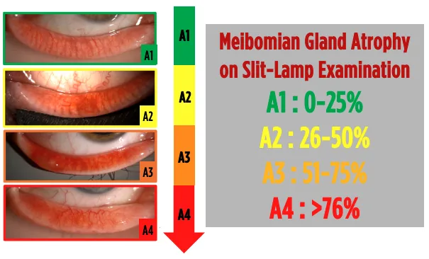
Meibomian Gland Atrophy
Careful examination of the everted eyelid can provide clinically useful information regarding meibomian gland morphology. With experience, the clinician can recognize gland shortening, irregularity, reduced gland density, and areas of visible glandular atrophy, and can semi-quantify the degree of structural involvement.
This clinical assessment provides an immediate estimation of structural gland disease. It does not replace infrared meibography, which offers a more complete and objective visualization of the entire meibomian gland architecture.
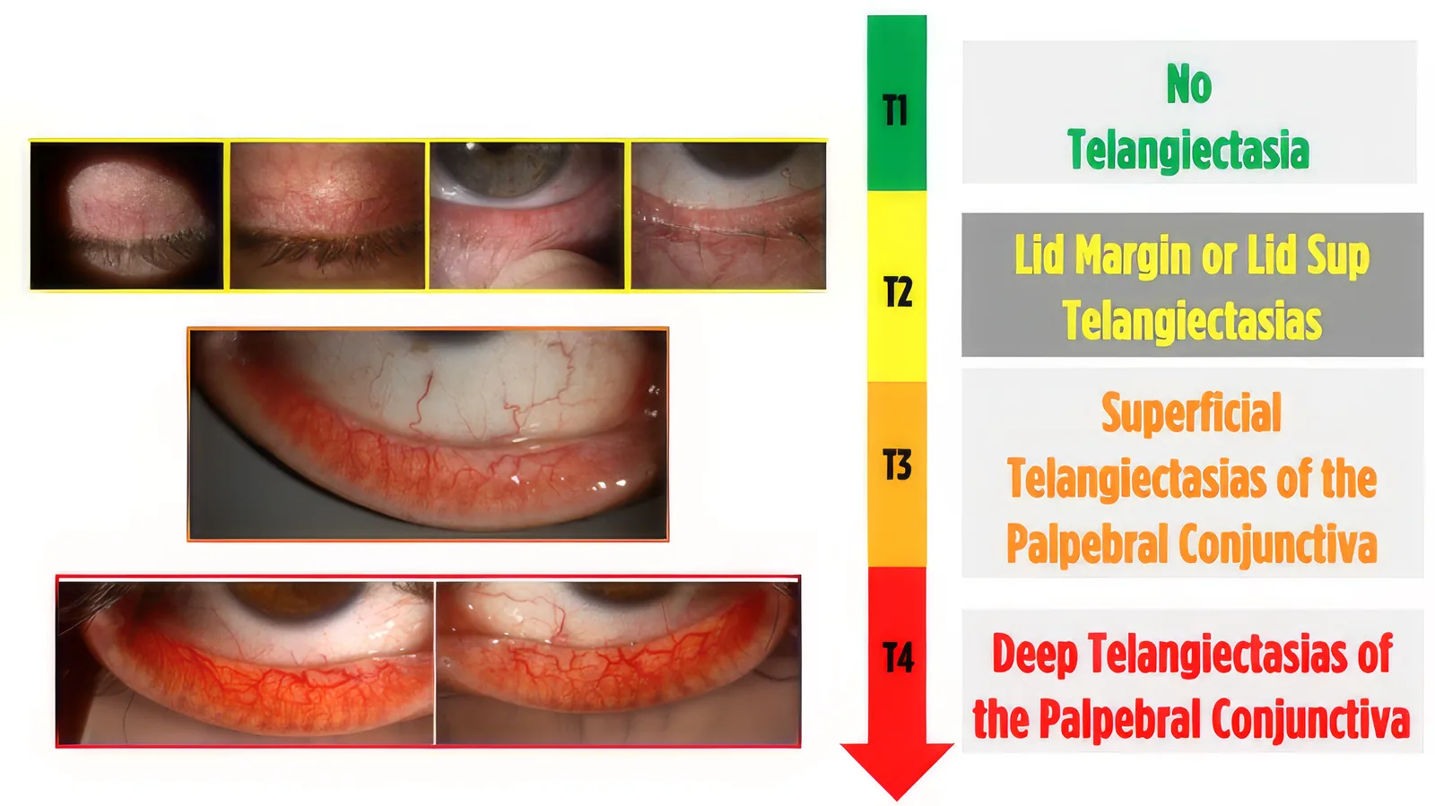
Telangiectatic Involvement
Abnormal vascularization of the lid margin is an important clinical feature of meibomian gland dysfunction, particularly in inflammatory disease and ocular rosacea.
Slit-lamp examination allows the clinician to assess and semi-quantify the presence, density, and extent of telangiectatic vessels along the lid margin and around the meibomian gland orifices. Progressive telangiectatic involvement may reflect an increasing inflammatory and vascular component of the disease and should be incorporated into the overall pathophysiological assessment.
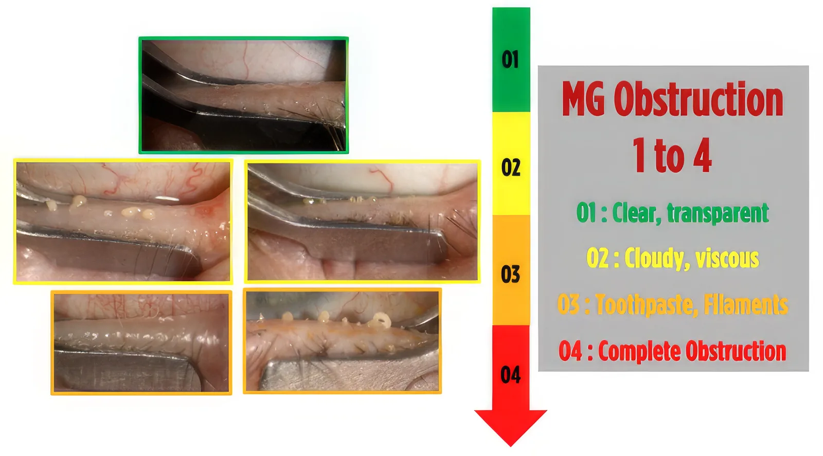
Meibomian Gland Obstruction
The appearance of the meibomian gland orifices provides direct information about obstructive MGD. Slit-lamp examination can identify orifice narrowing, plugging, capping, keratinization, and altered secretion at the gland openings.
With a systematic examination, the extent of gland orifice obstruction can be semi-quantified and contributes to the distinction between predominantly obstructive, inflammatory, and mixed forms of meibomian gland dysfunction. Functional evaluation by standardized meibomian gland expression provides complementary information regarding meibum quality and gland expressibility.
Tear Film Stability
Objective, non-invasive assessment of tear-film stability.
Tear film stability is a key component of dry eye assessment and reflects the ability of the tear film to remain intact between blinks.
BUT: Break-Up Time
Fluorescein Break-Up Time (BUT) is measured after instillation of fluorescein. The interval between a complete blink and the first appearance of a dry spot on the cornea is recorded.
A shortened BUT indicates tear-film instability and may reflect increased evaporation, altered lipid-layer function, or an abnormal ocular surface.
NIBUT: Non-Invasive Break-Up Time
Non-Invasive Break-Up Time (NIBUT) assesses tear-film stability without fluorescein.
Using corneal topography or dedicated tear-film imaging systems, NIBUT detects the first distortion or disruption of the reflected pattern on the corneal surface.
Because it does not disturb the tear film, NIBUT provides a more physiological and reproducible assessment of tear-film stability.
BUT and NIBUT are complementary functional tests that help quantify tear-film instability and document one of the major features of dry eye disease.
Ocular Surface Staining
Standardized assessment of ocular surface epithelial damage
Fluorescein and other vital dyes reveal epithelial damage that may not be visible during routine examination. The clinician should assess not only the presence of staining, but also its location, distribution, density, and severity.
Standardized grading systems, such as the Modified Oxford Grading Scale, improve reproducibility and allow changes to be documented over time. Staining patterns may also provide clues regarding the underlying mechanism of ocular surface disease.
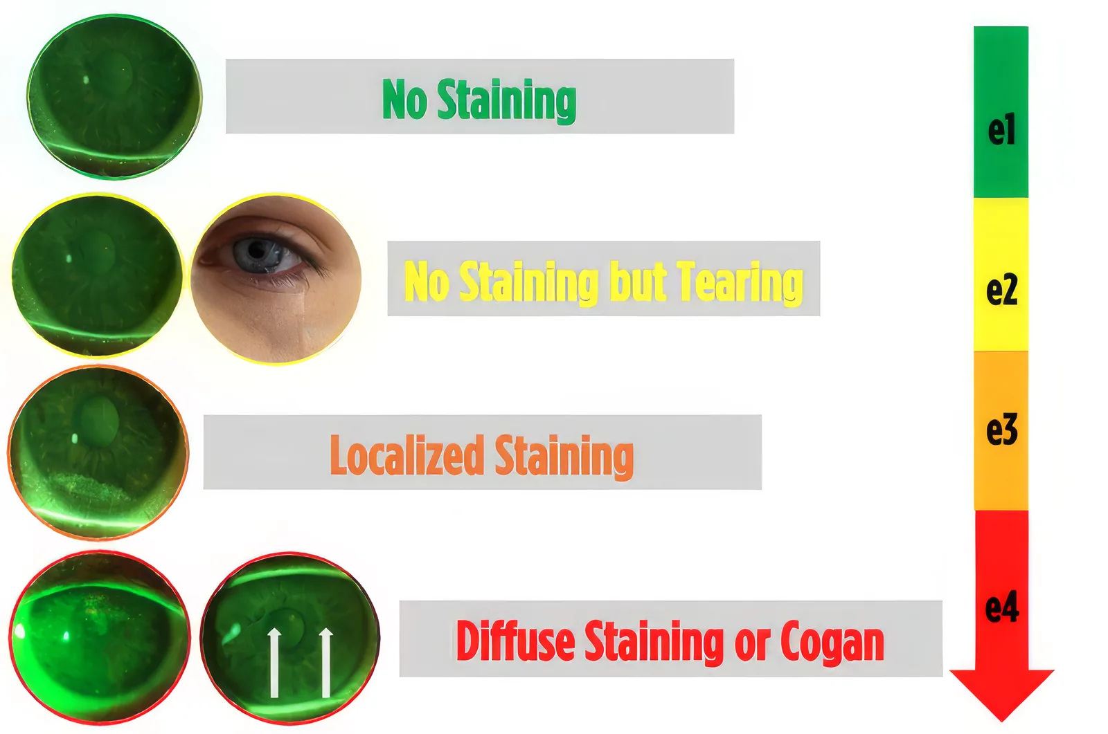
Ocular surface staining provides a simple and reproducible assessment of epithelial damage in dry eye disease.
Corneal Staining: Oxford Grading
Fluorescein staining allows direct visualization of corneal epithelial damage.
The Oxford Grading Scheme is a standardized method used to assess the severity of ocular surface staining. It compares the density of punctate staining observed on the ocular surface with a series of reference panels ranging from minimal or absent staining to severe epithelial damage.
The Oxford scale is commonly graded from 0 to 5, with increasing grades corresponding to increasing staining density.
It provides a practical method for:
- documenting baseline ocular surface damage;
- grading disease severity;
- comparing findings between visits;
- monitoring response to treatment.
Conjunctival Staining
Conjunctival staining complements corneal staining and provides a broader assessment of ocular surface involvement.
It is commonly evaluated using lissamine green or rose bengal, which highlight areas of epithelial compromise and mucin deficiency on the bulbar conjunctiva.
Assessment of both the cornea and conjunctiva provides a more complete picture of ocular surface epithelial damage.
When indicated, examination may also include the lid wiper, particularly when mechanical friction or lid wiper epitheliopathy is suspected.
Corneal and conjunctival staining are complementary tools for assessing the epithelial consequences of dry eye disease.
Meibography
Structural assessment of the meibomian glands.
Non-invasive infrared imaging provides direct visualization of meibomian gland morphology, allowing assessment of gland length, distortion, atrophy, and dropout.
Meibography helps determine the structural component of meibomian gland dysfunction and contributes to therapeutic decision-making.
Meibomian Gland Expression
Functional assessment of meibomian gland performance.
Standardized gland expression evaluates both meibum expressibility and secretion quality, providing essential information on the functional component of meibomian gland dysfunction.
Structural assessment by meibography and functional assessment by gland expression should be interpreted together.
Corneal OCT
High-resolution mapping of the corneal epithelium.
Corneal epithelial OCT provides an objective and reproducible assessment of epithelial thickness and distribution.
It may help identify epithelial remodeling and abnormalities associated with ocular surface disease and provides an additional objective parameter for longitudinal follow-up.
Complementary Investigations
Additional investigations may be selected according to the clinical presentation and suspected pathophysiological mechanisms.
Tear Osmolarity
Assessment of tear hyperosmolarity as an objective marker of altered tear-film and ocular-surface homeostasis.
Aqueous Tear Assessment
Schirmer testing and/or tear meniscus height may help identify an aqueous-deficient component, particularly in patients with severe dry eye or suspected systemic disease.
Blink Assessment
Assessment of blink frequency, blink completeness, interblink interval, and eyelid closure helps identify blink-related tear-film instability and exposure-related contributors to ocular surface disease.
Corneal Sensitivity
Corneal sensitivity testing should be considered in patients with severe ocular surface disease, reduced symptoms despite significant clinical signs, suspected neurotrophic keratopathy, or unexplained treatment failure.
In Vivo Confocal Microscopy
In selected complex cases, in vivo confocal microscopy can provide additional information regarding corneal nerves, inflammatory changes, and cellular alterations of the ocular surface.
Diagnostic Strategy
A multimodal assessment combines symptoms, careful slit-lamp examination, tear-film homeostasis, ocular surface damage, meibomian gland structure and function, and corneal epithelial changes to identify the predominant pathophysiological drivers and guide targeted treatment.
The first step is to confirm dry eye disease by identifying a loss of tear-film and/or ocular-surface homeostasis in a symptomatic patient.
The second step is to characterize the predominant pathophysiological drivers responsible for the disease in order to guide the selection and combination of treatments.
Accurate diagnosis identifies the disease. Mechanism-oriented assessment guides the interventional strategy.
A Clinical Overview Before Technology
Advanced diagnostic devices should complement, not replace, a careful clinical examination.
| Surface damage | ocular surface staining |
|---|---|
| Structural gland disease | visible gland abnormalities and atrophy |
| Inflammatory and vascular involvement | telangiectasia |
| Obstructive disease | gland orifice plugging and obstruction |
These findings provide the first pathophysiological orientation and help determine which objective investigations should be performed next.
The slit lamp provides the clinical picture. Advanced diagnostics refine and quantify it.
Accurate diagnosis is the foundation of personalized intervention.
Combining clinical examination, functional assessment and advanced imaging helps identify the mechanisms driving Dry Eye Disease and supports a targeted interventional strategy.