Corneal Epithelial Optical Coherence Tomography (eOCT) provides a non-invasive, objective, and reproducible pachymetric map of the corneal epithelium.
The corneal epithelium is a dynamic tissue continuously exposed to the tear film and the external environment. Chronic tear-film instability, inflammation, hyperosmolarity, and increased mechanical friction can progressively alter epithelial homeostasis and lead to measurable changes in corneal epithelial pachymetry.
Corneal epithelial pachymetric mapping provides an objective assessment of the structural impact of dry eye disease on the corneal surface.
Why Corneal Epithelial Pachymetry Matters
Slit-lamp examination and ocular surface staining identify epithelial damage, but they do not directly quantify the corneal epithelium.
Anterior-segment OCT adds another dimension by generating a micron-scale pachymetric map of the epithelium across the corneal surface.
It can provide information regarding:
- mean epithelial pachymetry;
- regional epithelial thinning;
- superior-inferior asymmetry;
- pachymetric variability;
- focal or diffuse epithelial abnormalities;
- changes in epithelial distribution over time.
In dry eye disease, central epithelial pachymetry may remain relatively preserved in some patients, whereas regional thinning, pachymetric asymmetry, and increased epithelial irregularity may become more apparent as ocular surface disease progresses.
From Tear-Film Dysfunction to Epithelial Damage
Dry eye disease exposes the corneal epithelium to repeated episodes of tear-film instability, hyperosmolarity, inflammation, and mechanical stress.
Over time, these mechanisms may disrupt normal epithelial homeostasis.
The resulting epithelial changes can range from subtle regional pachymetric abnormalities to more extensive epithelial thinning and irregularity.
Corneal epithelial OCT therefore provides a way to visualize and quantify the corneal consequences of chronic ocular surface dysfunction.
The tear film reflects instability.
Staining reveals epithelial damage.
Epithelial OCT quantifies its structural corneal impact.
WIDES Corneal Epithelial Pachymetry Grading
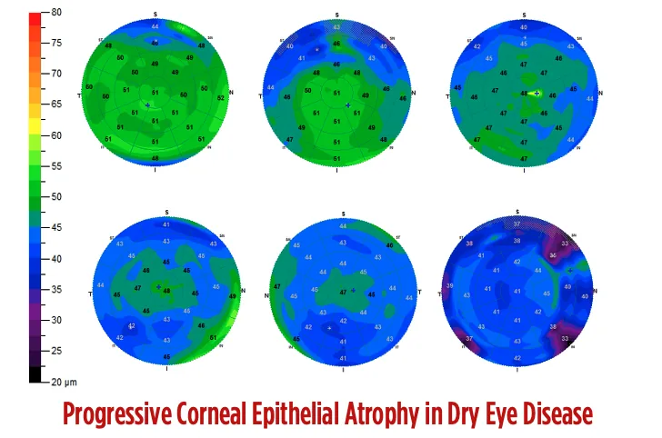
For clinical education, WIDES uses a four-stage visual grading approach to illustrate the progressive pachymetric changes observed in dry eye disease, from a preserved epithelial profile to advanced epithelial thinning and irregularity.
This grading is based on the overall epithelial pachymetric map and should always be interpreted together with ocular surface staining, tear-film stability, symptoms, and the complete clinical examination.
Preserved Epithelial Pachymetry
The epithelial pachymetric profile remains predominantly preserved, with no significant regional thinning or abnormal distribution.
Early Pachymetric Changes
Initial regional thinning or asymmetry becomes detectable, while most of the epithelial pachymetric map remains relatively preserved.
Moderate Epithelial Thinning
More extensive areas of reduced epithelial pachymetry and greater regional heterogeneity are visible, indicating a significant effect of ocular surface disease on epithelial homeostasis.
Severe Epithelial Thinning
Marked and extensive reduction in epithelial pachymetry with substantial disruption of the normal epithelial distribution, indicating severe corneal epithelial involvement.
The Importance of Pachymetric Distribution
Corneal epithelial OCT should not be interpreted from a single central value alone.
Dry eye-related epithelial abnormalities may be regional rather than uniformly distributed across the cornea.
Particular attention should therefore be paid to:
Regional pachymetric abnormalities may provide more clinically relevant information than central epithelial pachymetry alone.
The interpretation of the complete map is therefore essential.
Epithelial OCT and Ocular Surface Staining
Corneal epithelial OCT and ocular surface staining provide different but complementary information.
Ocular surface staining identifies areas of impaired epithelial integrity.
Corneal epithelial OCT objectively measures epithelial pachymetry and its spatial distribution.
A patient may therefore demonstrate pachymetric abnormalities even when staining is limited, while significant staining may occur without a proportional reduction in epithelial pachymetry.
The two examinations should therefore be interpreted together rather than considered interchangeable.
Staining shows where the epithelium is damaged.
OCT shows how its pachymetric architecture has changed.
An Objective Tool for Follow-Up
One of the major advantages of corneal epithelial OCT is its ability to provide a quantitative and reproducible pachymetric baseline.
Serial epithelial maps can help document changes in pachymetry and epithelial distribution over time and complement symptoms, staining, tear-film stability, and other clinical parameters during follow-up.
However, differences between OCT devices, acquisition protocols, segmentation algorithms, scan diameters, and peripheral measurement reliability must be considered when comparing examinations.
Whenever possible, longitudinal follow-up should therefore be performed using the same OCT device and the same acquisition protocol.
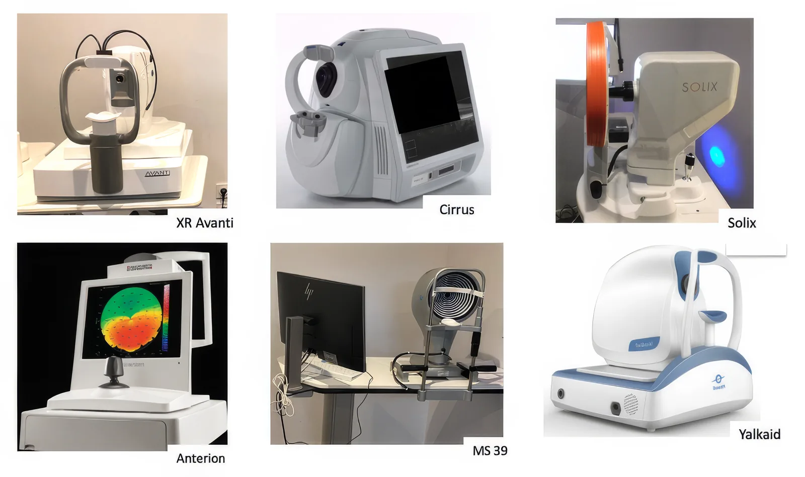
Corneal Epithelial OCT Devices
Several modern anterior-segment OCT platforms provide dedicated corneal epithelial pachymetric mapping.
Their performance may differ according to:
- scan diameter;
- axial resolution;
- epithelial segmentation algorithm;
- number of analyzed sectors;
- peripheral corneal coverage;
- pachymetric map reproducibility;
- ability to export and compare serial examinations.
For dry eye assessment, wide and reproducible epithelial pachymetric mapping is particularly useful because clinically relevant abnormalities may extend beyond the central cornea.
Epithelial OCT Is Not a Stand-Alone Dry Eye Test
Corneal epithelial OCT should not be interpreted in isolation.
There is normal biological variability in epithelial pachymetry, and abnormal pachymetric patterns may also occur in other corneal conditions.
The examination must therefore be integrated with:
Its principal value in Interventional Dry Eye is not simply to confirm the presence of dry eye disease, but to provide an objective measurement of its structural impact on the corneal epithelium.
Clinical Take-Home Message
Corneal epithelial OCT transforms epithelial involvement from a clinical impression into a measurable pachymetric parameter.
By mapping the pachymetry and spatial distribution of the corneal epithelium, it provides an objective assessment of the corneal impact of dry eye disease and complements tear-film evaluation, ocular surface staining, and meibomian gland assessment.
Tear-Film DysfunctionEpithelial StressPachymetric ChangesObjective OCT Mapping