Diagnostic meibomian gland expression provides a direct functional assessment of gland obstruction, meibum flow, expressibility, and secretion quality.
While meibography reveals gland structure, expression evaluates whether the remaining glands are able to deliver meibum, how easily secretion can be expressed, and how its quality and consistency have been altered.
A gland visible on meibography is not necessarily a functional gland.
For this reason, structural imaging and gland expression should be considered complementary components of the assessment of meibomian gland dysfunction (MGD).
Why gland expression matters
Meibomian gland dysfunction cannot be fully characterized by gland morphology alone.
Clinical studies, including work by Reiko Arita and colleagues, have shown the importance of combining gland morphology, meibum quality, and meibum expressibility, because structural gland loss and functional impairment provide related but distinct information.
Diagnostic expression allows the clinician to assess:
- gland obstruction;
- meibum flow;
- ease of expression;
- quality and transparency of the meibum;
- viscosity of the secretion;
- presence of particulate, filamentous, or inspissated material;
- partial or complete obstruction;
- differences between upper and lower eyelids.
Forceps-assisted gland expression
Direct visualization under the slit lamp
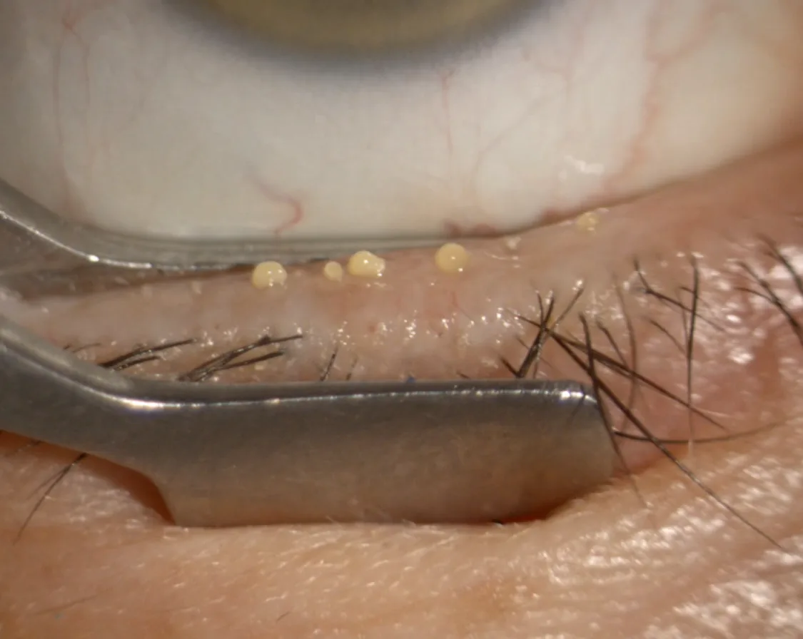
For WIDES clinical assessment, meibomian gland expression can be performed using a dedicated forceps, providing controlled support on the tarsal side of the eyelid while progressive compression is applied to the glands.
This technique allows the examiner to observe the gland orifices directly under slit-lamp magnification while assessing both the material expressed and the resistance encountered during expression.
The pressure should be progressive and controlled. Excessive force should not be used simply to obtain secretion, since the amount of pressure required is itself clinically informative.
Whenever practical, both the upper and lower eyelids should be assessed, as meibum quality and gland function may differ considerably between them.
What should be assessed?
Two complementary characteristics should always be considered during gland expression:
1. Meibum quality
Normal meibum is generally clear, transparent, and fluid.
With progressive MGD, the secretion may become:
Changes in secretion appearance provide important information about the degree of meibomian gland dysfunction.
2. Meibum expressibility
The clinician should simultaneously assess how easily secretion can be obtained.
Progressive dysfunction is associated with:
Meibum quality and expressibility should therefore always be interpreted together.
WIDES meibomian gland obstruction grading
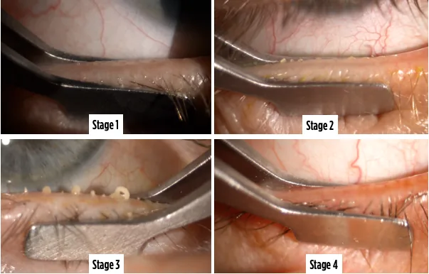
For clinical education and standardized documentation, WIDES uses a four-stage visual grading approach combining the appearance of expressed meibum with the degree of gland obstruction.
This simple visual classification is intended to provide a practical representation of the progressive functional deterioration of the meibomian glands.
Clear and fluid meibum is readily expressed with gentle compression.
The secretion remains essentially normal, with preserved meibum flow and minimal or no significant obstruction.
The secretion becomes cloudy and more viscous.
Expression remains possible but may require increased pressure, indicating early impairment of meibum flow and increasing gland obstruction.
Marked alteration in meibum consistency is present.
The expressed material may be thick, particulate, filamentous, or toothpaste-like, and substantially greater pressure may be required to evacuate the gland.
This stage corresponds to advanced obstructive dysfunction with markedly altered meibum flow.
No meibum can be expressed despite adequate controlled compression.
The gland is functionally non-expressible.
Structural assessment by meibography is particularly important at this stage to distinguish severe obstruction of a structurally preserved gland from advanced glandular atrophy.
Structure and function must be interpreted together
Meibography and gland expression answer two different questions.
How much glandular tissue remains and what does it look like?
How obstructed are the glands, can meibum still flow, and what is the quality of the secretion?
Structural preservation does not necessarily guarantee normal gland function, and structural gland loss alone does not provide a complete assessment of meibomian gland dysfunction.
Meibography shows the structure.
Expression reveals the function.
Obstruction versus atrophy
This distinction is particularly important in Interventional Dry Eye.
A patient may present with:
The combination of meibography and gland expression therefore provides a more meaningful assessment than either examination alone.
Why this matters for treatment
Identifying the relative contribution of obstruction, altered meibum quality, inflammation, and structural gland loss is essential when selecting an interventional strategy.
Gland expression should therefore be interpreted together with:
This multimodal approach helps distinguish patients with predominantly obstructive, inflammatory, atrophic, or mixed MGD phenotypes.
The goal is not simply to diagnose MGD, but to identify the predominant mechanism responsible for gland dysfunction.
An objective baseline for follow-up
The initial gland expression grade should be recorded whenever possible.
Repeating gland expression after treatment provides a simple clinical method for documenting changes in:
- meibum clarity;
- viscosity;
- meibum flow;
- expressibility;
- number of functioning glands;
- degree of obstruction.
Whenever longitudinal comparisons are made, the same eyelid regions and a similar expression technique should be used to improve reproducibility.
Clinical take-home message
Meibomian gland expression is the functional counterpart of meibography.
It determines how obstructed the glands are, whether meibum can still flow, and how the quality and expressibility of the secretion have been altered.
Combining gland expression with meibography provides a more complete characterization of meibomian gland dysfunction and helps identify the predominant pathophysiological mechanism before treatment.