Meibography is a non-invasive imaging technique that allows direct visualization of the meibomian glands within the tarsal plates.
Using infrared light, it reveals gland morphology that cannot be fully assessed by routine slit-lamp examination and provides a structural complement to the functional information obtained by meibomian gland expression.
Meibography does not simply show whether glands are present or absent. It provides a morphological map of meibomian gland disease.
Why meibography matters
Meibomian gland dysfunction (MGD) is one of the major contributors to evaporative dry eye disease.
Clinical examination of the lid margin can reveal obstruction, telangiectasia, inflammation, or abnormal meibum, but it does not provide direct visualization of the entire glandular architecture.
Infrared meibography allows clinicians to assess:
- gland length;
- gland width and density;
- tortuosity;
- distortion;
- shortening and truncation;
- areas of gland dropout;
- partial or advanced gland atrophy;
- differences between the upper and lower eyelids.
These morphological changes provide important information about the structural component of meibomian gland dysfunction.
Examine all four eyelids
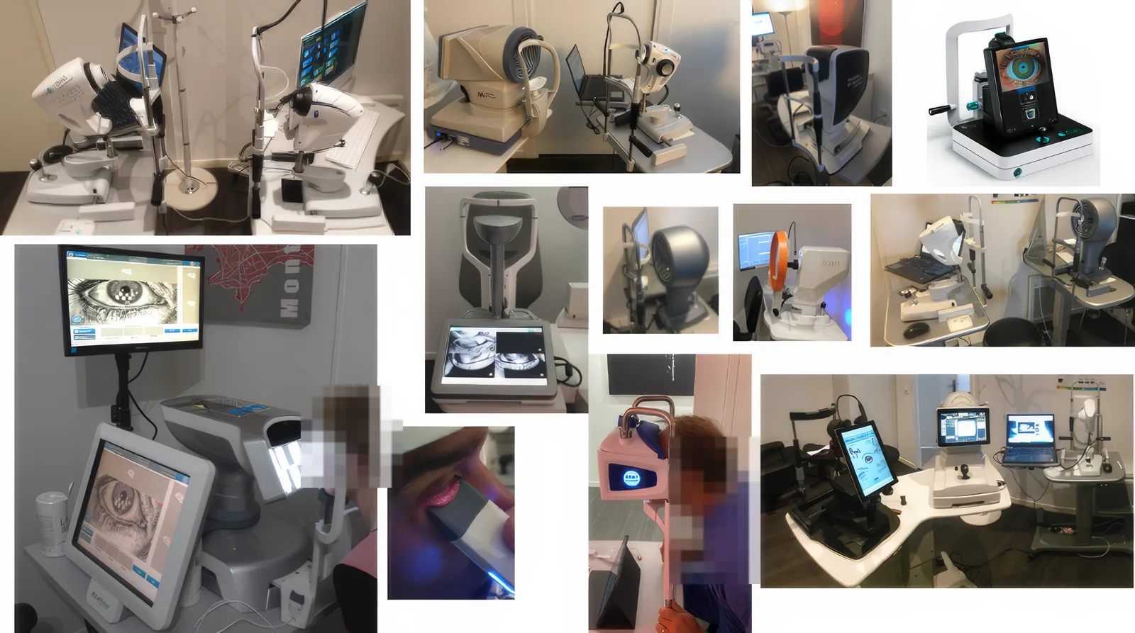
A complete meibographic assessment should include both the upper and lower eyelids of both eyes.
The morphology and degree of gland loss may differ substantially between eyelids. Examination of only one or two eyelids may therefore provide an incomplete picture of the patient's glandular status.
Comparison of the four eyelids provides a more comprehensive assessment of:
This four-eyelid approach is particularly useful for identifying asymmetric or localized disease and for establishing a reliable structural baseline.
What should be analyzed?
Meibography should not be reduced to a simple percentage of gland loss.
The clinician should evaluate the overall gland architecture, including gland length, regularity, distortion, tortuosity, density, shortening, and areas of partial or complete dropout.
Some morphological abnormalities may be present before advanced gland loss becomes evident, whereas severe disease is characterized by progressive shortening, disruption of normal architecture, and increasing areas in which glandular tissue is no longer visible.
No single morphological feature should be interpreted in isolation.
Meibography is most informative when integrated with functional gland assessment and the overall ocular surface examination.
Meibomian gland atrophy grading
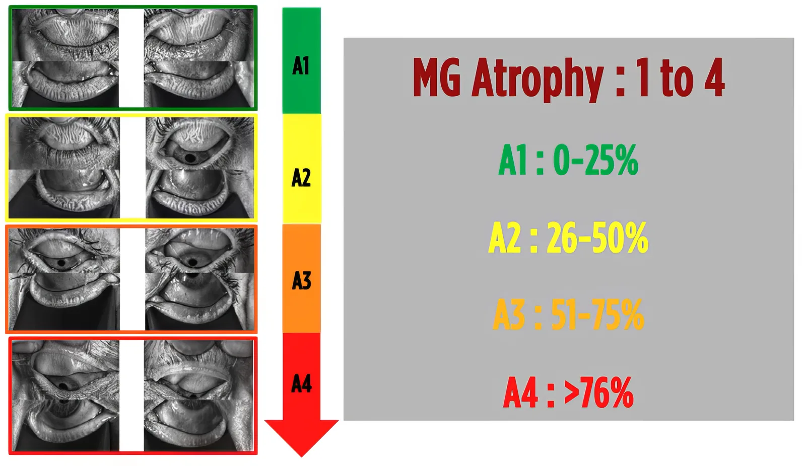
For clinical education, WIDES illustrates the progression of meibomian gland atrophy using a four-stage visual grading system, based on the estimated percentage of gland loss within the tarsal area and incorporating the assessment of all four eyelids. The grading should be interpreted together with gland morphology, including shortening, distortion, irregularity, and loss of normal gland architecture.
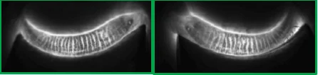
Predominantly preserved glandular architecture, with long and well-defined glands. Minimal focal shortening, distortion, or dropout may be present.
Partial gland loss becomes clearly visible, with increasing shortening, distortion, irregularity, and reduction in gland density.
Marked reduction in gland number, length, and density, with extensive areas of dropout and substantial disruption of normal glandular architecture.
Extensive meibomian gland dropout with severe loss of normal glandular architecture and only limited residual gland tissue.
Structure is not function
Meibography and meibomian gland expression provide complementary information.
A gland that remains visible on meibography may nevertheless be functionally impaired.
Conversely, structural abnormalities do not always correlate directly with the severity of symptoms.
For this reason, structural imaging should always be interpreted together with:
Meibography shows the gland. Expression shows what the gland can still produce.
Telangiectatic involvement & ocular rosacea
Looking beyond gland atrophy
Meibomian gland dysfunction is not solely an obstructive or atrophic process.
Inflammatory and vascular abnormalities, particularly in ocular rosacea, may coexist with altered meibomian secretion and progressive structural gland changes.
Lid-margin telangiectasia is an important clinical marker of this inflammatory and vascular component.
Assessment of telangiectatic involvement may therefore provide additional information regarding the mechanisms contributing to meibomian gland dysfunction and may influence the therapeutic strategy.
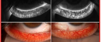
WIDES telangiectatic involvement grading
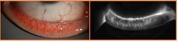
For clinical documentation and education, WIDES uses a four-stage visual approach to illustrate progressive telangiectatic involvement associated with meibomian gland disease.
This visual grading should always be interpreted together with direct slit-lamp examination of the lid margin and the complete ocular surface assessment.
No significant abnormal vascular changes associated with the meibomian gland area.
Limited vascular changes with focal telangiectatic involvement.
More extensive telangiectatic changes associated with increasing glandular abnormalities.
Marked vascular and inflammatory involvement associated with extensive structural gland abnormalities.
Meibography and treatment selection
Meibography is not a treatment-selection tool on its own.
Its role is to identify the structural substrate of meibomian gland disease and contribute to a broader pathophysiological assessment.
When combined with gland expression, tear-film analysis, lid-margin examination, ocular surface staining, and other diagnostic findings, meibography can help identify patients with predominantly:
This distinction is central to the WIDES approach because different pathophysiological patterns may require different therapeutic strategies or combinations of treatments.
Longitudinal follow-up
Meibography provides a permanent visual record of meibomian gland morphology.
Serial imaging may be useful for documenting structural evolution over time and identifying progressive gland loss.
However, short-term clinical improvement after treatment should not necessarily be expected to produce visible restoration of glands that are already severely atrophic.
Treatment efficacy should therefore be assessed using a combination of:
rather than changes in meibography alone.
Clinical take-home message
Meibography reveals the structural component of meibomian gland disease.
Its greatest clinical value comes from combining the morphological information provided by infrared imaging with functional gland expression and assessment of the tear film, ocular surface, eyelids, and inflammatory environment.